
News
Kunststoff
Unternehmen
INNOTEQ 2023: Nachhaltige Swissness für die MEM-Industrie

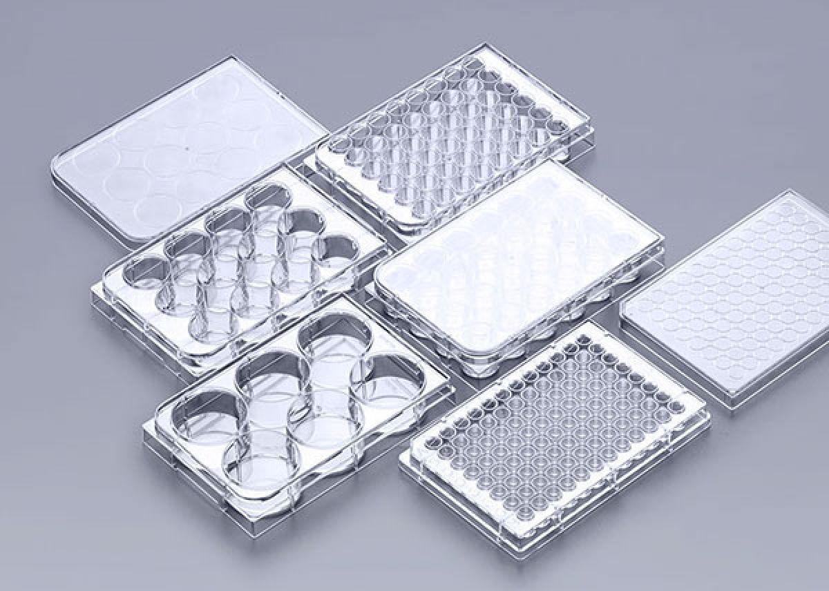
HEK293 cells are one of the most commonly used tools in protein expression, viral packaging, and functional studies. However, many researchers often encounter challenges like low efficiency and unsatisfactory protein expression levels during transfection. This usually stems not from a single error, but from the cumulative effect of subtle deviations in multiple key steps. This article will focus on 5 crucial yet easily overlooked points to help you systematically improve HEK293 cell transfection outcomes and protein yield.
Key Point 1: Ensure Optimal Cell State and Passage Number
Core Principle: Use healthy cells in the logarithmic growth phase with a low passage number.
Problem: Over-passaged cells (e.g., beyond passage 50) may experience genotypic and phenotypic drift, leading to reduced growth rate and transfection sensitivity. Cells in poor condition or in the plateau phase show significantly lower transfection efficiency.
Solutions:
Regularly thaw cells from a low-passage stock, avoiding excessive continuous subculturing.
Ensure cells are in the logarithmic growth phase at the time of transfection, with viability ᢗ%.
After seeding, allow cells to recover in the incubator for at least 24 hours to ensure complete attachment and optimal growth condition.
Key Point 2: Precisely Optimize Seeding Density and Transfection Timing
Core Principle: The cell confluence at the time of transfection is critical for success.
Problem: Excessively high cell density leads to contact inhibition and nutrient competition; excessively low density means insufficient factors secreted by cells that promote DNA binding, and unstable growth status.
Solutions:
For HEK293 cells, a confluence between 70% and 90% at transfection is typically the optimal window.
Perform pilot experiments to determine the optimal seeding density. For example, seed cells in a 24-well plate 24 hours in advance at a density of 2-5×10^5 cells/mL per well.
Key Point 3: Use High-Purity, High-Quality Plasmid DNA
Core Principle: The purity and concentration of the plasmid directly determine the formation efficiency of transfection complexes.
Problem: Plasmids contaminated with endotoxins, proteins, or RNA can interfere with the formation of transfection complexes and cause cytotoxicity, leading to low efficiency and low expression.
Solutions:
Prepare plasmids using endotoxin-removal kits (e.g., based on anion-exchange columns).
Check plasmid concentration and purity using a spectrophotometer, ensuring an A260/A280 ratio between 1.8 and 1.9.
For critical experiments, agarose gel electrophoresis is recommended to confirm the supercoiled structure and absence of degradation.
Key Point 4: Select the Appropriate Transfection Reagent and Meticulously Optimize the Ratio
Core Principle: There is no "universal" reagent; optimizing the DNA-to-transfection reagent ratio is crucial.
Problem: Polyethylenimine (PEI) is a common reagent for HEK293 cell transfection, but its efficiency highly depends on the DNA (μg) to PEI (μl) ratio. Improper ratios lead to uneven complex size, excessive toxicity, or low encapsulation efficiency.
Solutions:
Perform gradient experiments testing different DNA:PEI ratios (e.g., 1:1, 1:2, 1:3, 1:4).
Protocol Tip: Dilute DNA and PEI separately in serum-free medium (e.g., Opti-MEM), let them sit for 5 minutes before mixing, then mix and incubate the mixture at room temperature for 15-20 minutes to form stable transfection complexes.
Key Point 5: Post-Transfection Culture Management and Cell Health Monitoring
Core Principle: Post-transfection handling also affects the final protein yield.
Problem: Transfection reagents have cytotoxicity; prolonged exposure can affect cell health; improper medium change procedures can disturb the cells.
Solutions:
Replace the medium with fresh complete culture medium 4-6 hours after transfection to remove the transfection reagent and replenish nutrients.
If the expression period is long (>48 hours), consider a partial medium change at 24 hours post-transfection.
Closely monitor cell morphology and health. If significant cytotoxicity is observed, try shortening the co-incubation time of the complexes with the cells.
Regularly perform mycoplasma testing. Mycoplasma contamination is a hidden culprit behind poor cell health and unstable experimental results.
Summary
Improving protein expression in HEK293 cells is a systematic project. By strictly controlling these five key points—cell state, seeding density, plasmid quality, transfection reagent ratio, and post-transfection management—you can significantly enhance the reproducibility and success rate of your experiments.